Diaphragm paralysis: a case report
Keywords:
Diaphragm paralysis, dyspnoea, respiratory failure, diaphragmatic ultrasoundAbstract
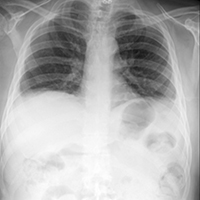
Introduction. Bilateral paralysis of the diaphragm may be an idiopathic clinical condition or associated with several diseases such as trauma, surgery, viral infections, neurologic disorders. The diaphragm is the main respiratory muscle. It is a cupoliform muscle-tendon structure, innervated bilaterally by phrenic nerve, which originates from C3-C5 nerve roots. Diaphragmatic paralysis is a clinical disorder that generates hypoventilation and basal pulmonary atelectasis, predisposing to hypercapnic respiratory failure. The clinic manifestations mimic cardio-respiratory pathologies, therefore often misdiagnosticated. Case presentation. A 55-year-old man with a previous C6-7 traumatic fracture, referred multiple accesses to the emergency room for acute nocturnal dyspnoea, treated with antibiotic therapy, diuretic therapy and long-term oxygen therapy, without beneficial effects. He referred to our pulmonary clinic for evaluation of persistent and worsening orthopnoea due to unknown cause for about 2 years. Clinical examination, respiratory functional tests and diaphragm ultrasound revealed a strong suspicion of diaphragmatic deficit, confirmed by electromyography. Conclusions. The patient accesses to the emergency room numerous times and the clinical frame have been always oriented towards a cardio-respiratory origin. From the onset of the symptom to the respiratory evaluation, about 2.5 years have passed. The manifestation of clear orthopnoea has addressed the functional respiratory study towards a more thorough diaphragmatic evaluation assessed by ultrasound.
References
Podnar S. Idiopathic phrenic neuropathies: A case series and review of the literature. Muscle Nerve 2015;52:986-92.
Fromageot C, Lofaso F, Annane D, Falaize L, Lejaille M, Clair B, et al. Supine fall in lung volumes in the assessment of diaphragmatic weakness in neuromuscular disorders. Arch Phys Med Rehabil 2001;82:123-8.
Ko MA, Darling GE. Acquired paralysis of the diaphragm. Thorac Surg Clin 2009;19:501-10.
Caruso P, Albuquerque ALP de, Santana PV, Cardenas LZ, Ferreira JG, Prina E, et al. Diagnostic methods to assess inspiratory and expiratory muscle strength. J Bras Pneumol 2015;41:110-23.
Kokatnur L, Rudrappa M. Diaphragmatic palsy. Diseases 2018;6:16.
Ricoy J, Rodríguez-Núñez N, Álvarez-Dobaño JM, Toubes ME, Riveiro V, Valdés L. Diaphragmatic dysfunction. Pulmonology 2019;25:223-35.
Published
Issue
Section
License
Mattioli 1885 has chosen to apply the Creative Commons Attribution NonCommercial 4.0 International License (CC BY-NC 4.0) to all manuscripts to be published.
How to Cite


