A single-center comparative study of lung ultrasound versus chest computed tomography during the COVID-19 era
Keywords:
COVID-19, lung ultrasonography, computed tomography, consolidation, B-linesAbstract
Background: Lung ultrasound (LUS) is a bedside imaging tool that has proven useful in identifying and assessing the severity of pulmonary pathology. The aim of this study was to determine LUS patterns, their clinical significance, and how they compare to CT findings in hospitalized patients with coronavirus infection.
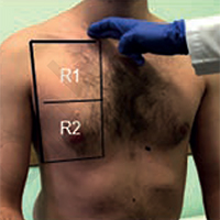
Methods: This observational study included 62 patients (33 men, age 59.3±15.9 years), hospitalized with pneumonia due to COVID-19, who underwent chest CT and bedside LUS on the day of admission. The CT images were analyzed by chest radiographers who calculated a CT visual score based on the expansion and distribution of ground-glass opacities and consolidations. The LUS score was calculated according to the presence, distribution, and severity of anomalies.
Results: All patients had CT findings suggestive of bilateral COVID-19 pneumonia, with an average visual scoring of 8.1±2.9%. LUS identified 4 different abnormalities, with bilateral distribution (mean LUS score: 26.4±6.7), focal areas of non-confluent B lines, diffuse confluent B lines, small sub-pleural micro consolidations with pleural line irregularities, and large parenchymal consolidations with air bronchograms. LUS score was significantly correlated with CT visual scoring (rho = 0.70; p<0.001). Correlation analysis of the CT and LUS severity scores showed good interclass correlation (ICC) (ICC =0.71; 95% confidence interval (CI): 0.52–0.83; p<0.001). Logistic regression was used to determine the cut-off value of ≥27 (area under the curve: 0.97; 95% CI: 90-99; sensitivity 88.5% and specificity 97%) of the LUS severity score that represented severe and critical pulmonary involvement on chest CT (CT: 3-4).
Conclusion: When combined with clinical data, LUS can provide a potent diagnostic aid in patients with suspected COVID-19 pneumonia, reflecting CT findings.
References
Volpicelli G, Gargani L. Sonographic signs and patterns of COVID-19 pneumonia. Ultrasound J 2020;12:22.
Volpicelli G, Lamorte A, Villén T. What’s new in lung ultrasound during the COVID-19 pandemic. Intensive Care Med 2020;46:1445-8.
Sultan LR, Sehgal CM. A Review of early experience in lung ultrasound in the diagnosis and management of COVID-19. Ultrasound Med Biol 2020;46:2530-45.
World Health Organization. Clinical management of severe acute respiratory infection (SARI) when COVID-19 disease is suspected: interim guidance, 13 March 2020. Accessed: March 13, 2020. Available from: https://apps.who.int/iris/handle/10665/331446
Nouvenne A, Zani MD, Milanese G, Parise A, Baciarello M, Bignami EG, et al. Lung ultrasound in COVID-19 pneumonia: Correlations with chest CT on hospital admission. Respiration 2020;99:617-24.
Zhang R, Ouyang H, Fu L, Wang S, Han J, Huang K, et al. CT features of SARS-CoV-2 pneumonia according to clinical presentation: a retrospective analysis of 120 consecutive patients from Wuhan city. Eur Radiol 2020;30:4417-26.
Li K, Fang Y, Li W, Pan C, Qin P, Zhong Y, et al. CT image visual quantitative evaluation and clinical classification of coronavirus disease (COVID-19). Eur Radiol 2020;30:4407–16.
Bao C, Liu X, Zhang H, Li Y, Liu J. Coronavirus disease 2019 (COVID-19) CT findings: A systematic review and meta-analysis. J Am Coll Radiol 2020;17:701–9.
Tung-Chen Y, Martí de Gracia M, Díez-Tascón A, Alonso-González R, Agudo-Fernández S, Parra-Gordo ML, et al. Correlation between chest computed tomography and lung ultrasonography in patients with coronavirus disease 2019 (COVID-19). Ultrasound Med Biol 2020;46:2918-26.
Bouhemad B, Mongodi S, Via G, Rouquette I. Ultrasound for ‘lung monitoring’ of ventilated patients. Anesthesiology 2015;122:437–47.
Long C, Xu H, Shen Q, Zhang X, Fan B, Wang C, et al. Diagnosis of the coronavirus disease (COVID-19): rRT-PCR or CT? Eur J Radiol 2020;126:108961.
Ai T, Yang Z, Hou H, Zhan C, Chen C, Lv W, et al. Correlation of chest CT and RT-PCR testing in coronavirus disease 2019 (COVID-19) in China: a report of 1014 cases. Radiology 2020;296:E32-40.
Chung M, Bernheim A, Mei X, Zhang N, Huang M, Zeng X, et al. CT Imaging features of 2019 novel coronavirus (2019-nCoV). Radiology 2020;295:202-7.
Bernheim A, Mei X, Huang M, Yang Y, Fayad ZA, Zhang N, et al. Chest CT findings in coronavirus disease-19 (COVID-19): relationship to duration of infection. Radiology 2020;295:200463.
Colombi D, Bodini FC, Petrini M, Maffi G, Morelli N, Milanese G, et al. Well-aerated lung on admitting chest CT to predict adverse outcome in COVID-19 pneumonia. Radiology 2020;296:E86-96.
Rubin GD, Ryerson CJ, Haramati LB, Sverzellati N, Kanne JP, Raoof S, et al. The role of chest imaging in patient management during the COVID-19 pandemic: A multinational consensus statement from the Fleischner Society. Chest 2020;158:106-16.
Allinovi M, Parise A, Giacalone M, Amerio A, Delsante M, Odone A, et al. Lung ultrasound may support diagnosis and monitoring of COVID-19 pneumonia. Ultrasound Med Biol 2020;46:2908-17.
Gargani L, Soliman-Aboumarie H, Volpicelli G, Corradi F, Pastore MC, Cameli M. Why, when, and how to use lung ultrasound during the COVID-19 pandemic: enthusiasm and caution. Eur Hear J Cardiovasc Imaging 2020;21:941-8.
Piscaglia F, Stefanini F, Cantisani V, Sidhu PS, Barr R, Berzigotti A, et al. Benefits, open questions and challenges of the use of ultrasound in the COVID-19 pandemic era. The views of a panel of worldwide international experts. Ultraschall Med 2020;41:228–36.
Soldati G, Smargiassi A, Inchingolo R, Buonsenso D, Perrone T, Briganti DF, et al. Proposal for international standardization of the use of lung ultrasound for COVID-19 patients; a simple, quantitative, reproducible method. J Ultrasound Med 2020;39:1413-9.
Poggiali E, Dacrema A, Bastoni D, Tinelli V, Demichele E, Mateo Ramos P, et al. Can lung US help critical care clinicians in the early diagnosis of novel coronavirus (COVID-19) pneumonia? Radiology 2020;295:E6.
Lieveld AWE, Kok B, Schuit FH, Azijli K, Heijmans J, van Laarhoven A, et al. Diagnosing COVID-19 pneumonia in a pandemic setting: Lung ultrasound versus CT (LUVCT) - a multicentre, prospective, observational study. ERJ Open Res 2020;6:00539-2020.
McDermott C, Daly J, Carley S. Combatting COVID-19: is ultrasound an important piece in the diagnostic puzzle? Emerg Med J 2020;37:644-9.
Published
Issue
Section
License
Mattioli 1885 has chosen to apply the Creative Commons Attribution NonCommercial 4.0 International License (CC BY-NC 4.0) to all manuscripts to be published.
How to Cite