Diaphragmatic excursion by ultrasound: reference values for the normal population; a cross-sectional study in Egypt
Keywords:
diaphragmatic ultrasound, diaphragmatic excursion, diaphragmatic motion, M-mode ultrasound, reference values, normal valuesAbstract
Background: Measurement of diaphragmatic motion by ultrasound is being utilized in different aspects of clinical practice. Defining reference values of the diaphragmatic excursion is important to identify those with diaphragmatic motion abnormalities. This study aimed to define the normal range of diaphragmatic motion (reference values) by M-mode ultrasound for the normal population.
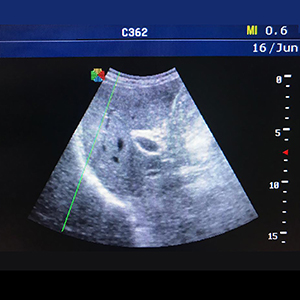
Methods: Healthy volunteers were included in this study. Those with comorbidities, skeletal deformity, acute or chronic respiratory illness were excluded. Diaphragmatic ultrasound in the supine position was performed using a low frequency probe. The B-mode was applied for diaphragmatic identification, and the M-mode was employed for the recording of the amplitude of diaphragm contraction during quiet breathing, deep breathing and sniffing.
Results: The study included 757 healthy subjects [478 men (63.14%) and 279 women (36.86%)] with normal spirometry and negative history of previous or current respiratory illness. Their mean age and BMI were 45.17 ±14.84 years and 29.36±19.68 (kg/m2). The mean right hemidiaphragmatic excursion was 2.32±0.54, 5.54±1.26 and 2.90±0.63 for quiet breathing, deep breathing and sniffing respectively, while the left hemidiaphragmatic excursion was 2.35±0.54, 5.30±1.21 and 2.97±0.56 cm for quiet breathing, deep breathing and sniffing respectively. There was a statistically significant difference between right and left diaphragmatic excursion among all studied subjects. The ratio of right to left diaphragmatic excursion during quiet breathing was (1.009±0.19); maximum 181% and minimum 28%. Only 19 cases showed a right to left ratio of less than 50% (5 men and 14 women). The diaphragmatic excursion was higher in males than females. There was a significant difference in diaphragmatic excursion among age groups. Age, sex and BMI significantly affected the diaphragmatic motion.
Conclusions: Diaphragmatic excursion values presented in this study can be used as reference values to detect diaphragmatic dysfunction in clinical practice. Diaphragmatic motion is affected by several factors including age, sex and body mass index.
References
Lloyd T, Tang YM, Benson MD, King S. Diaphragmatic paralysis: The use of M mode ultrasound for diagnosis in adults. Spinal Cord 2006;44:505-8.
Gierada DS, Curtin JJ, Erickson SJ, Prost RW, Strandt JA, Goodman LR. Diaphragmatic motion: Fast gradient-recalled-echo MR imaging in healthy subjects. Radiology 1995;194:879-84.
Houston JG, Fleet M, Cowan MD, McMillan NC. Comparison of ultrasound with fluoroscopy in the Assessment of suspected hemidiaphragmatic movement abnormality. Clin Radiol 1995;50:95–8.
Li G, Wei J, Huang H, Gaebler CP, Yuan A, Deasy JO. Automatic assessment of average diaphragm motion trajectory from 4DCT images through machine learning. Biomed Phys Eng Express 2015;1:045015.
Epelman M, Navarro OM, Daneman A, Miller SF. M-mode sonography of diaphragmatic motion: Description of technique and experience in 278 pediatric patients. Pediatr Radiol 2005;35:661–7.
Testa A, Soldati G, Giannuzzi R, Berardi S, Portale G, Gentiloni Silveri N. Ultrasound M-Mode assessment of diaphragmatic kinetics by anterior transverse scanning in healthy subjects. Ultrasound Med Biol 2011;37:44–52.
Pellegrino R, Viegi G, Brusasco V, Crapo RO, Burgos F, Casaburi R, et al. Interpretative strategies for lung function tests. Eur Respir J 2005;26:948–68.
Boussuges A, Rives S, Finance J, Brégeon F. Assessment of diaphragmatic function by ultrasonography: Current approach and perspectives. World J Clin Cases 2020;8:2408-24.
Gerscovich EO, Cronan M, McGahan JR, Jain K, Jones CD, McDonald C. Ultrasonographic evaluation of diaphragmatic motion. J Ultrasound Med 2001;20:597-604.
McCool FD, Tzelepis GE. Dysfunction of the diaphragm. N Engl J Med 2012;366:932-42.
Boussuges A, Finance J, Chaumet G, Brégeon F. Diaphragmatic motion recorded by M-mode ultrasonography: limits of normality. ERJ Open Res 2021;7:00714-2020.
Scarlata S, Mancini D, Laudisio A, Benigni A, Antonelli Incalzi R. Reproducibility and clinical correlates of supine diaphragmatic motion measured by M-Mode ultrasonography in healthy volunteers. Respiration 2018;96:259-66.
Kantarci F, Mihmanli I, Demirel MK, Harmanci K, Akman C, Aydogan F, et al. Normal diaphragmatic motion and the effects of body composition: Determination with M-mode sonography. J Ultrasound Med 2004;23:255–60.
Vieira Santana P, Zumpano Cardenas L, Luis Pereira de Albuquerque A, Roberto Ribeiro de Carvalho C, Caruso P. Diaphragmatic ultrasound: a review of its methodological aspects and clinical uses. J Bras Pneumol 2020;46:e20200064.
Sarwal A, Walker FO, Cartwright MS. Neuromuscular ultrasound for evaluation of the diaphragm. Muscle Nerve 201;47:319.
Umbrello M, Formenti P. Ultrasonographic assessment of diaphragm function in critically ill subjects. Respir Care 2016;61:542-55.
Published
Issue
Section
License
Copyright (c) 2022 The Author(s)

This work is licensed under a Creative Commons Attribution-NonCommercial 4.0 International License.
Mattioli 1885 has chosen to apply the Creative Commons Attribution NonCommercial 4.0 International License (CC BY-NC 4.0) to all manuscripts to be published.
How to Cite


